Wednesday, July 13, 2016

Wow! Beautiful Bridges.

I love it when we get a "Wow!" from patients after seeing their new smile for the first time. Here are two bridges we recently cemented. Can you tell which teeth are part of the bridge?

BRIDGE 1:


This patient's natural dentition has traces of white spots. The bridge matches this pattern wonderfully.
This bridge is made up of the three middle teeth in the picture. The middle tooth is the fake one (pontic).

 
BRIDGE 2:


This bridge is the last 4 teeth (to the right) in the picture. The missing tooth/ pontic is second from the last. There is one support tooth behind the missing tooth and two supporting teeth in front. This patient was ecstatic with the results.


Saturday, June 11, 2016

Rampant Decay Before and After


Rampant decay, meaning cavities everywhere, is a challenging problem to fix. Causes of the disease and time and cost of treatment are all obstacles to overcome. Many times, without definitive action to correct, these patients will end up losing most if not all their teeth. 


Before: Notice decay near gumline and in between teeth. Areas of white/opaque enamel and loss of translucency indicates decalcification and the beginning stages of decay.


After: Day of delivery. Crowned/ bridged all upper teeth. One canine tooth had been extracted and is a pontic (fake tooth) on a bridge. Patient was extremely happy with results. 


Monday, May 30, 2016

"Doc, My Filling Came Out." (w/ PICTURES)

When someone visits our office and reports that a "filling came out," in most cases, it is actually part of the tooth that broke and not the filling.

Having been an ENGINEER before becoming a dentist, I look for tooth problems created by chewing and grinding forces. I look for clues that teeth might be weak and need reinforcement. One of my goals is to evaluate structural integrity of teeth, and to help every patient keep and enjoy their teeth for a lifetime.

The below photos show some broken teeth. Documentation helps to calibrate my understanding of when a tooth is truly at risk, so I can make better recommendations in the future. As they say, a picture is worth a thousand words. Enjoy!


CASE 1: "BUT IT DOESN'T HURT"


Yellow A: Corner of tooth broke and missing.
Red B: Worn spot on adjacent crown. Metal showing through.
Green C: Shiny wear spots on filling.
Despite multiple clues indicating occlusal problems, the patient was not interested in treating tooth at this time. The patient stated it was not hurting them. 

Yellow A: Original break.
Red B: New break causing pain.
Green C: Tongue.
The second break (Red B) was much more severe, and it was unknown at this point if the tooth could be saved. We needed to numb the patient and remove the tooth piece to see the extent of the break. 

Yellow A: Very deep break.
To restore the tooth, it required root canal therapy and gum surgery in addition to the originally needed crown procedures. The patients was sorry they didn't get just the crown when that would have prevented this more extensive treatment.

Outside view of broken piece.

Inside view of broken piece.


CASE 2: SURPRISE!


Yellow A: Wear patterns on teeth. Enamel layer worn off. Softer dentin showing through.
This tooth was weaker than I expected. Before we were able to replace the filling, the tooth broke. I did not give enough urgency to these wear patterns.

Yellow A: This outlines shows the extent of missing tooth.

Saliva is filling the hole left by the missing tooth piece.

Outside view, broken tooth.

Inside view, broken piece.
Notice how sharp the break is at the bottom.


CASE 3: BACK TOOTH BREAK


Back, tongue-side cusp of back tooth is broken.


Numbed patient and removed broken piece of tooth.
No root canal therapy was needed. Crown procedures restored function and strength.


Outside view, broken piece. Note gum attachment at bottom of piece.


CASE 4: NON-RESTORABLE BREAK


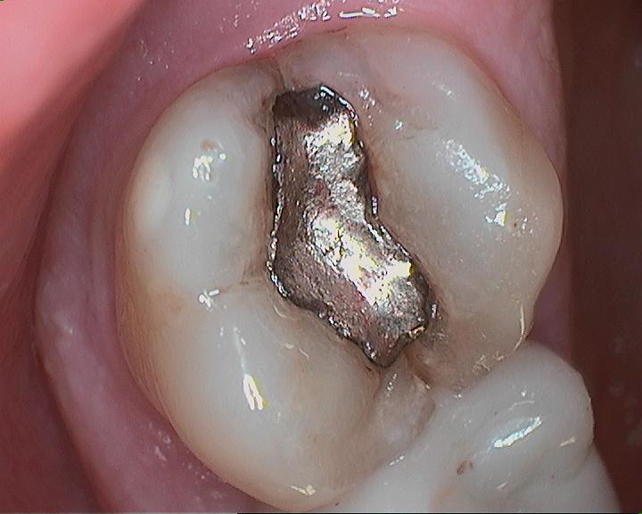
Top back tooth presents with crack under old filling.

Filling was removed without needing to drill. Older silver fillings are not bonded and are only mechanically retained.

Depth of crack prevented ability to restore tooth. Tooth was extracted.


Sunday, December 27, 2015

How Clean Is Clean?

A patient recently commented to me about how "clean" her teeth felt after our hygienist had finished. I was pleased that the patient enjoyed the newly polished and smooth texture of her dentition; however, I also saw an opportunity for a little education.

Part of TOOTH ANATOMY includes leftover vestiges of development called dentinal tubules. These microscopic pores provide the perfect hiding place for bacteria and yeast. This fact makes it practically impossible to mechanically remove all microbes from a tooth's surface. This is why many times I will recommend a chemical therapy as an adjunct to regular hygiene maintenance.

DID YOU KNOW?... The human body contains more bacterial cells than human cells. Consider the mouth, gut, skin, etc. In fact many of these bacterial cells are actually beneficial to us.


Thursday, September 24, 2015

Tired of Your Denture? Have REAL Teeth Again!

New Materials and Techniques Allow Dentists to Do New Procedures

When a person is missing all or most of their teeth, the options for replacing them become limited. Nowadays dentists can use implants and stronger materials to restore full arches of teeth.  A lot of patients are choosing this option and the satisfaction rate is very high.

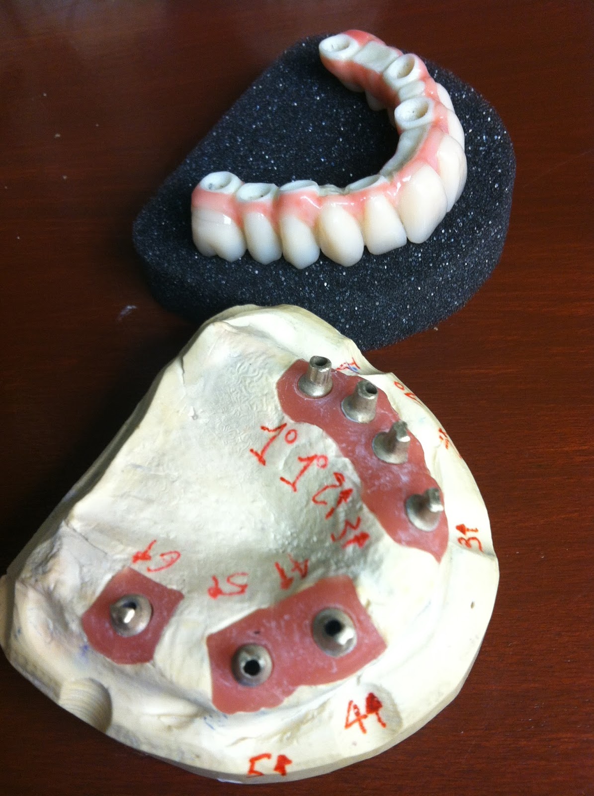
The below pictures are of a case recently delivered by Carmichael Dental Care. This is a "fixed hybrid" Trinia prosthesis on 7 implants. A minimum of 4 implants is required for this type of treatment.


Abutment Visible


Occlusal View


Mounted View 


Smile View


This patients was so happy to have his old, natural looking teeth back!... and they didn't come in and out of his mouth. He said he was going to go eat a steak that night.

Dentistry has a lot to offer and is a great way to help people improve their quality of life.

If you're in Alabama and have questions, feel free to call the office. We'd be happy to schedule a free consultation with you. Our number is 334-270-9924 or you can visit our website www.myALdentist.com


Friday, July 17, 2015

WARNING: GRAPHIC - Wisdom Tooth Surgery Photos

The following photographs are from a wisdom tooth surgery. 
Proceed with caution if you have a low tolerance for graphic pictures.
**********


Pre-op radiograph showing bony impaction and distoangular orientation.
**********


Pre-op intraoral photograph and planned incision.
**********


Gingival (gum) flap elevation exposing bone and tooth.
**********


Bur removal of bone in front, on the side and behind tooth.
**********


Tooth removed.
**********


Sutures (stitches) in place. 
**********


Surgery performed under oral conscious sedation.  Patient reports a very pleasant in-office experience but, as expected, was quite sore the next day.  Strong pain relievers and rest were able to manage the discomfort and kickstart recovery.


Tuesday, June 2, 2015

Twelve Days To My Wedding! Enamel Microabrasion

This patient is getting married in 12 days. What are his options?
How can the discoloration of the front teeth be reduced?


Before enamel microabrasion.
 
 
Enamel microabrasion is the process of applying an acid/pumice slurry that chemically and mechanically removes a thin layer of enamel and with it, any surface stains or discoloration. 
 
Results vary depending on the depth of the discoloration. Not everyone is a candidate.
 
As the reader can see from the below photo, this patient (and his fiancé) were very pleased.
 
 
After enamel microabrasion.
 
Another success story from Carmichael Dental Care.

Friday, May 15, 2015

Another Beautiful Case


When lab work gets delivered to our office, we check it for quality.  We strive to ensure the best possible end result to our patients.  Here is another beautiful case by our high quality lab partner.  

Visit us at www.myALdentist.com.

Friday, May 8, 2015

What is a Dry Socket?


Here is a patient-submitted photograph of their dry socket following a tooth extraction.  Notice the dark area behind the last tooth.  

When the healing blood clot that typically forms over an extraction site gets removed, for one reason or another, the bony socket where the tooth used to be is known as a "dry socket". 

These are usually very sore and painful.  The healing cannot very well be accelerated, but the aching can be managed with the application of a medicated dressing by the surgeon.  

Increased risk factors for dry socket includes:  SMOKING, diabetes, vigorous liquid swishing too soon after surgery, drinking carbonated beverages after surgery, eating hard or sharp food, applying negative pressure as when sucking through a straw, etc.

Avoid these activities after surgery.


Friday, March 13, 2015

Interesting Radiographs for you Dentistry Nerds

How mandibles heal on their own after almost 20 years.


Panoramic Radiograph 1996


Panoramic Radiograph 2015


Friday, January 30, 2015

Esthetics of Translucency


The patient loved her new crowns! 
Her old ones (porcelain-fused-to-metal) were very opaque so that no light could shine through.  This greatly reduced esthetic appeal, and they looked fake.  These new ones are made of different material and allow for light to show from behind - very similar to tooth enamel.  The perceived glow greatly enhances esthetic appeal.  Thank you Tosch Laboratory!

Saturday, January 17, 2015

Antibiotics for Prosthetic Joint Infection Prophylaxis


Do you have an artificial joint?

The Journal of the American Dental Association (JADA) published this month that patients with prosthetic joints no longer need to promedicate prior to dental treatment.

A panel has judged that current best evidence fails to demonstrate a link between dental procedures and prosthetic joint infection (PJI).  On the other hand, the risk of antibiotic resistance, adverse drug reactions, and cost associated with prescribing antibiotics for PJI prophylaxis is much greater.

To read the JADA article, click here.

Wednesday, December 31, 2014

A Lifetime of Wear: Full Arch Dentistry

What years of grinding and upper porcelain crowns can do to lower teeth.


Worn lower dentition with interdental spaces (spaces between teeth). 


Diagnostic wax-up showing proper height to width ratio of teeth and interdental spaces closed.


Treatment plan includes a 4 unit bridge (#18-21) and 2 implant crowns (#29, 30).

Dentistry can greatly improve quality of life for many people. It's rewarding to be a part of that process, and I enjoy applying Engineering principles to Dentistry.  

Stay tuned for more pictures.